Keywords: Cone beam computed tomography (CBCT), Temporomandibular joint disorder, TMD, TMJ, 3D imaging, orofacial pain, neck pain, migraine, and headache.
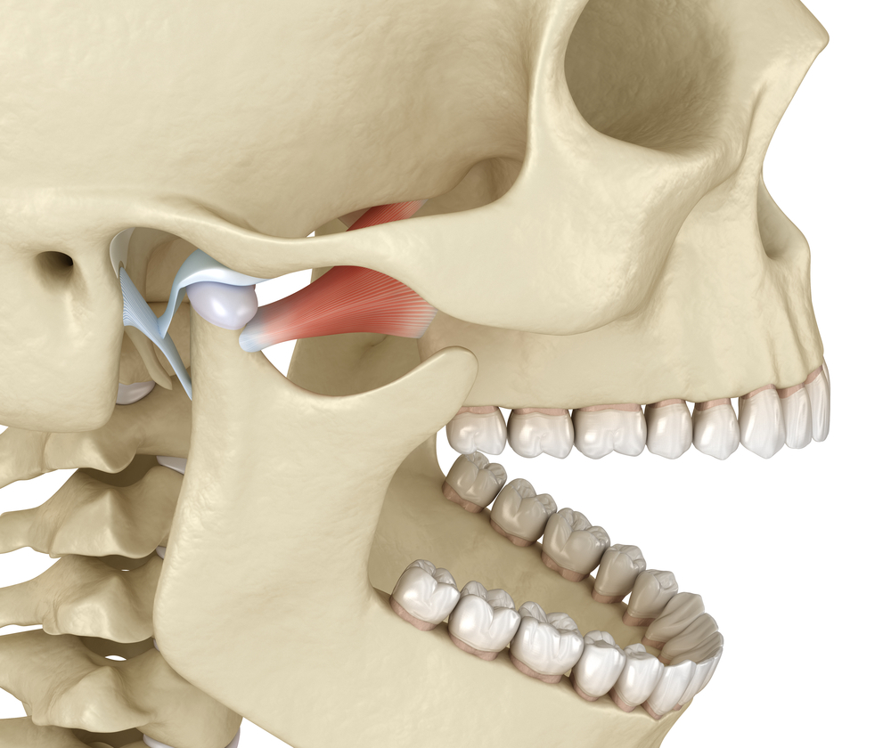
Cone beam computed tomography (CBCT) is a technique being used more often in dentomaxillofacial imaging. The cone-shaped X-ray beam centered on a two dimensional (2D) detector produces a series of 2D images. The reconstruction of the images into 3 dimensional (3D) images is done using the modified Feldkamp algorithm. This allows for an accurate 3D look into the patient’s temporomandibular joint (TMJ) and more importantly, an accurate diagnosis of temporomandibular joint disorder (TMD). The scan time of a 3D cone beam is 8.9 seconds and it is 1/10th the radiation of a medical CT scan. At Head Pain Institute, we don’t just treat your symptoms; we diagnose and treat the cause of your discomfort to enable you to live your life more comfortably. If you or someone you know suffers from TMD, headaches, migraines, and or facial pain, please schedule your initial consultation with Dr. Teruel or visit us at www.headpaininstitute.com for more information.
Krishnamoorthy B1, Mamatha N, Kumar VA. 1Department of Oral Medicine and Radiology, ITS-Centre of Dental Sciences and Research, Delhi-Meerut Road, Muradnagar, Ghaziabad, Uttar Pradesh, India.